In predicting a stroke’s toll, location matters, but so do connections

Each year, roughly 666,000 Americans survive a stroke, and for them, the aftermath can be hard to predict. Some stroke patients have difficulty speaking or grasp for words that do not come. Some suffer problems with vision, balance or mobility. Some are addled by attention, memory and other cognitive deficits that can range from subtle to severe.
To glean what kinds of disabilities a patient will probably face, neurologists have long looked at the location of the lesion a stroke leaves behind — on a brain scan, the darkened site where cells have died off. But when a system as complex as the human brain comes under attack, pinpointing the injury’s whereabouts isn’t always a very good guide.
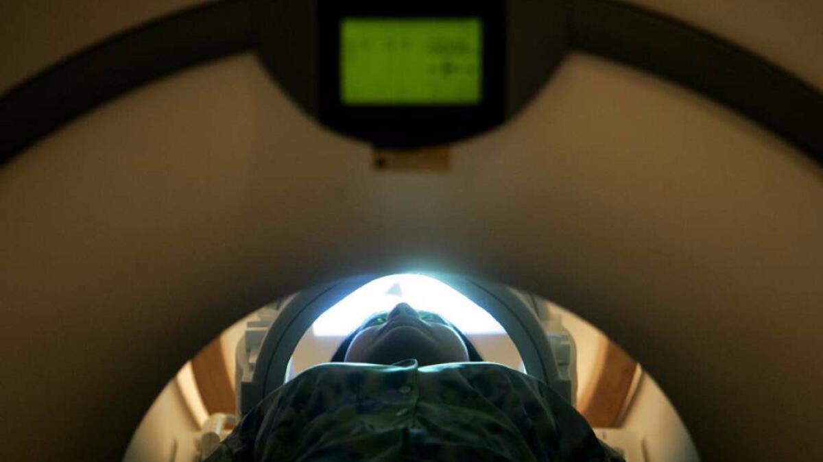
New research takes a key step toward arming physicians with better tools to predict the challenges a patient faces after a stroke. Within two weeks of 130 patients’ first stroke, researchers from Washington University School of Medicine put 132 stroke patients through a battery of brain scans that gauged not only the exact location of cellular damage but how communications traffic within their brain differed from that of healthy controls of similar age. Computer-based learning systems digested the resulting mass of data in a bid to discern patterns that could improve prediction.
Published Monday in the Proceedings of the National Academies of Science, the study underscores that, for many strokes, the location of lesions matters less than the disruptions a stroke causes in the flow of signals between the brain’s two hemispheres and within the sometimes far-flung networks that collectively govern human behavior.
That is especially true when predicting disabilities involving visual and verbal memory, where a stroke patient has trouble recognizing faces, places or things and attaching names to them. These are skills that require input from and coordination among a wide range of brain structures, and the authors of the latest paper found that when communications traffic slowed across the brain’s two hemispheres and among the structures that make up the brain’s key networks, it was a likely bet that these were the challenges that would bedevil a stroke patient.
The location of a stroke’s physical damage was actually a good predictor of sensorimotor deficits, including partial blindness or weakness or paralysis affecting one side of the body. When disrupted blood flow caused brain cells to die off in the cortical areas directly responsible for vision or motor function, or in the bundles of connective fiber that lash them together, the researchers found that such patients had a high probability of those disabilities.
Post-stroke language problems occupied an interesting middle ground. A doctor might predict language problems with about equal accuracy whether she located a stroke lesion in the brain’s highly specialized language region or she observed disrupted communications within the brain. Post-stroke language problems were seen in patients who had impaired communications within the brain’s specialized language areas, which lie usually in the brain’s left hemisphere. But they were also seen in stroke patients whose showed a loss of connection between the right and left hemisphere.
Language disorders, the researchers said, cannot only arise from “pure disruption of language processing.” They can also result when regions responsible for language lose their feed from support processes across the brain’s hemispheric divide.
While comparisons among stroke patients helped glean patterns of disabilities post-stroke, comparisons between stroke patients on the one hand and healthy control subjects on the other showed that connectivity problems between the brain’s two hemispheres are a hallmark problem for stroke survivors.
“Far and away the greatest change we see is reduced interhemispheric activity, reduced inhemispheric connections,” said lead author Joshua Sarfaty Siegel, a neuroscientist at Washington University in St. Louis. “Generally, no matter where the lesion is, there’s this phenomenon of reduced interhemispheric connectivity.”
Follow me on Twitter @LATMelissaHealy and “like” Los Angeles Times Science & Health on Facebook.
MORE IN SCIENCE
A ‘slow catastrophe’ unfolds as the golden age of antibiotics comes to an end
Science proves it: Girl Scouts really do make the world a better place
The damage wrought by acidic oceans hurts more than marine life and lasts longer than you think


